Measuring proliferation in breast cancer

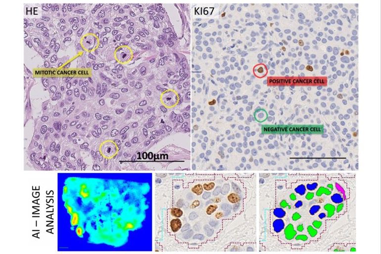
Breast cancer affects more than 2 million individuals globally each year. An important part of the diagnostic process is to assess proliferation in the tumour tissue, which is an indicator of how quickly the cancer is growing. It can give insight into the likely outcome of a patient and whether or not the patient should receive chemotherapy. This prognostic evaluation is currently performed by a visual assessment which involves counting cancer cells (see examples in the figure below).
The aim of this work package is to develop and validate artificial intelligence (AI) to develop tools to measure, standardize and improve reproducibility and accuracy of this assessment. The AI tool shall be developed for use as a decision-support tool by a pathologist. It’s use aims to improve turnaround time and accuracy of current evaluation methods, and allow for the reallocation of pathologist time to more complex aspects of diagnosis.
Currently, the project has worked on assessing the reproducibility of different AI methods and traditional manual counting methods, as well as assessing the prognostic validity of such decision-support tools. Results suggest, that AI may be equal to or better than current methods.

Figure: In the top left image, mitotic cancer cells are marked and counted in HE stained breast tumour tissue. In the top right, breast tumour tissue is stained to detect the proliferation marker Ki67. Cells that are positive for this marker appear brown, whilst negative cells appear blue. The higher the % of brown (positive) cells, the poorer the prognosis. In the bottom panel is an example of how AI methods can be used to identify highly proliferative regions and these can then be counted automatically.