Kidney fibrosis
Any chronic kidney disease will develop fibrosis, an increase of connective tissue, over time. Fibrosis is a key prognostic marker. Quantification of fibrosis is therefore crucial to assess prognosis and to guide therapy.
Kidney fibrosis is routinely evaluated by a rough estimate of the extend of fibrotic areas (slight,moderate, severe). This is difficult in routinely hematoxylin-eosin stained sections, because fibrosis is only weakly stained. The image below illustrates this: There is severe fibrosis in the left part and slight fibrosis in the right part.

Therefore special stains, usually trichrome or sirius red, are used. These stains highlight fibrosis blue (trichrome, left image below) or red (sirius red, right image below).

Sirius red is our chosen stain for evaluation of interstitial fibrosis, because it has a high signal to noise ratio which makes it suitable for automatic image analysis.
The aims of this study are:
To quantify interstitial fibrosis by conventional image analysis in sirius red stained slides from kidney biopsies (below)

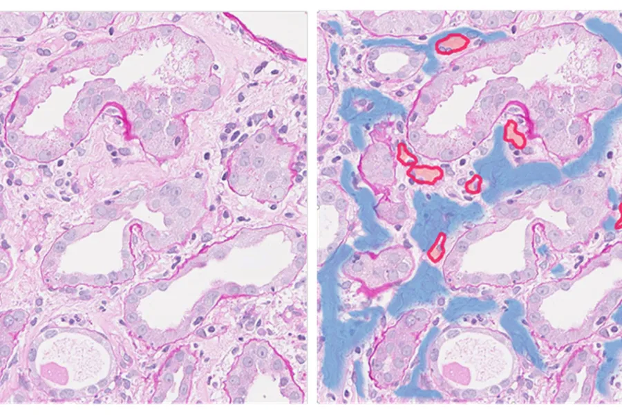
To use mark-up images from conventional image analysis to train a machine learning network to detect and quantify kidney fibrosis in routinely hematoxylin-eosin stained slides from kidney biopsies.