Ki-67 in neuroendocrine tumours
This work package is aimed to find an automated way for measuring Ki-67 index in neuroendocrine tumors of the gastrointestinal tract. The research will give insight into a number of image analysis applications and their advantages and disadvantages by describing the faced challenges and suggesting solutions.
The objectives are:
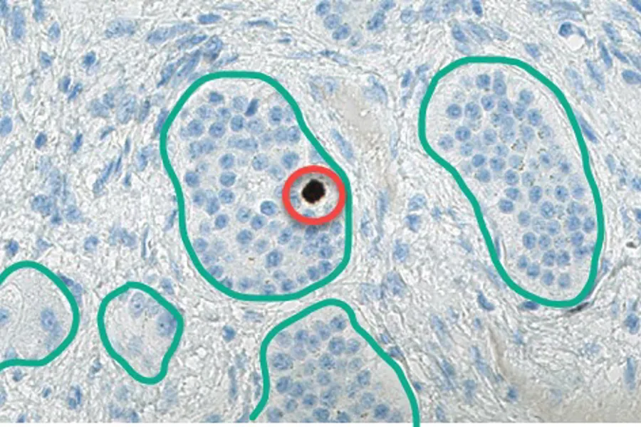
To establish the ground truth

Here has a pathologist manually counted negative (= pink) and positive (=cyan) tumour cells. This manual count is considered the "ground truth".
To perform conventional image analysis

This conventional image analysis application marks negative (=blue) and positive (=brown) tumour cells. It is easy to see that the demarcation of individual tumour cells does not work well.
To train and test a machine learning system

The trained algorithm performs very well in counting negative (= red) and positive (= green) tumour cells.
Finally, to compare different analysis systems with the ground truth.